External View Of Brain / Lab Practical Exam - Biology 255 with Carlson at Luther ... - It provides access to an atlas and to images in axial planes, allowing the user to learn.
External View Of Brain / Lab Practical Exam - Biology 255 with Carlson at Luther ... - It provides access to an atlas and to images in axial planes, allowing the user to learn.. It is divided into two cerebral hemispheres. It resulted in 5 different zones of separation, 1−5. In an experiment, extract of brain tissue was subjected to differential centrifugation. A medical illustration of the human brain from 'quain's elements of anatomy, eighth edition, vol.ii' (by william sharpey md, lld, frs l&e, allen thomson, md, lld, frs l&e, and edward albert schafer) depicts the right half of. Brain, coronal section, basal ganglia, close view, gross.
What are the structural and functional units of the brain? A medical illustration of the human brain from 'quain's elements of anatomy, eighth edition, vol.ii' (by william sharpey md, lld, frs l&e, allen thomson, md, lld, frs l&e, and edward albert schafer) depicts the right half of. View and manage file attachments for this page. The brain is also divided into several lobes: The cerebral cortex is an outer layer of grey matter, covering the core of white matter.
Sheep Brain Exam at Columbia University - StudyBlue from classconnection.s3.amazonaws.com It is the most complex organ in a vertebrate's body. It is made of billions of interconnected neurones and has different regions that carry out different functions. Any of a long list of feelings a person can have such as joy, anger and love. The figure can be manipulated with certain interaction functions to display more detailed information. Brain and spinal cord, gross. A few useful tools to. Use the map below to take a tour of the regions in the brain and learn what they control in. Brain, coronal section, basal ganglia, close view, gross.
It is the most complex organ in a vertebrate's body.
Try some of these examples. It provides access to an atlas and to images in axial planes, allowing the user to learn. The brain is also divided into several lobes: Moreover, brainnet viewer draws the brain surface, nodes and edges in sequence and displays brain networks in multiple views, as required by the user. Brain, coronal section, basal ganglia, close view, gross. How to view anatomical labels. Brain and spinal cord, gross. Physiological method of choice to record the electrical activity. • the frontal lobes are responsible for problem solving and judgment and motor function. A major aspect of the complexity of nervous systems relates to their intricate morphology, especially the effective connectivity may be viewed as the union of structural and functional connectivity, as it describes networks of directional. One promising strategy for enabling such a system, denoted neuralnanorobotics may also enable a b/ci with controlled connectivity between neural activity and external data storage and processing, via the. The interface combines electroencephalography (eeg) to record brain signals and transcranial magnetic stimulation (tms) to deliver information noninvasively to the brain. When viewed from above, a large groove (interhemispheric fissure) separates the brain into left and right halves.
It separates the temporal lobe from the frontal lobe. Although such an approach has been remarkably productive, it ignores the alternative possibility that brain functions are mainly … How to view anatomical labels. It is the most complex organ in a vertebrate's body. Study the same and answer the questions that follows:
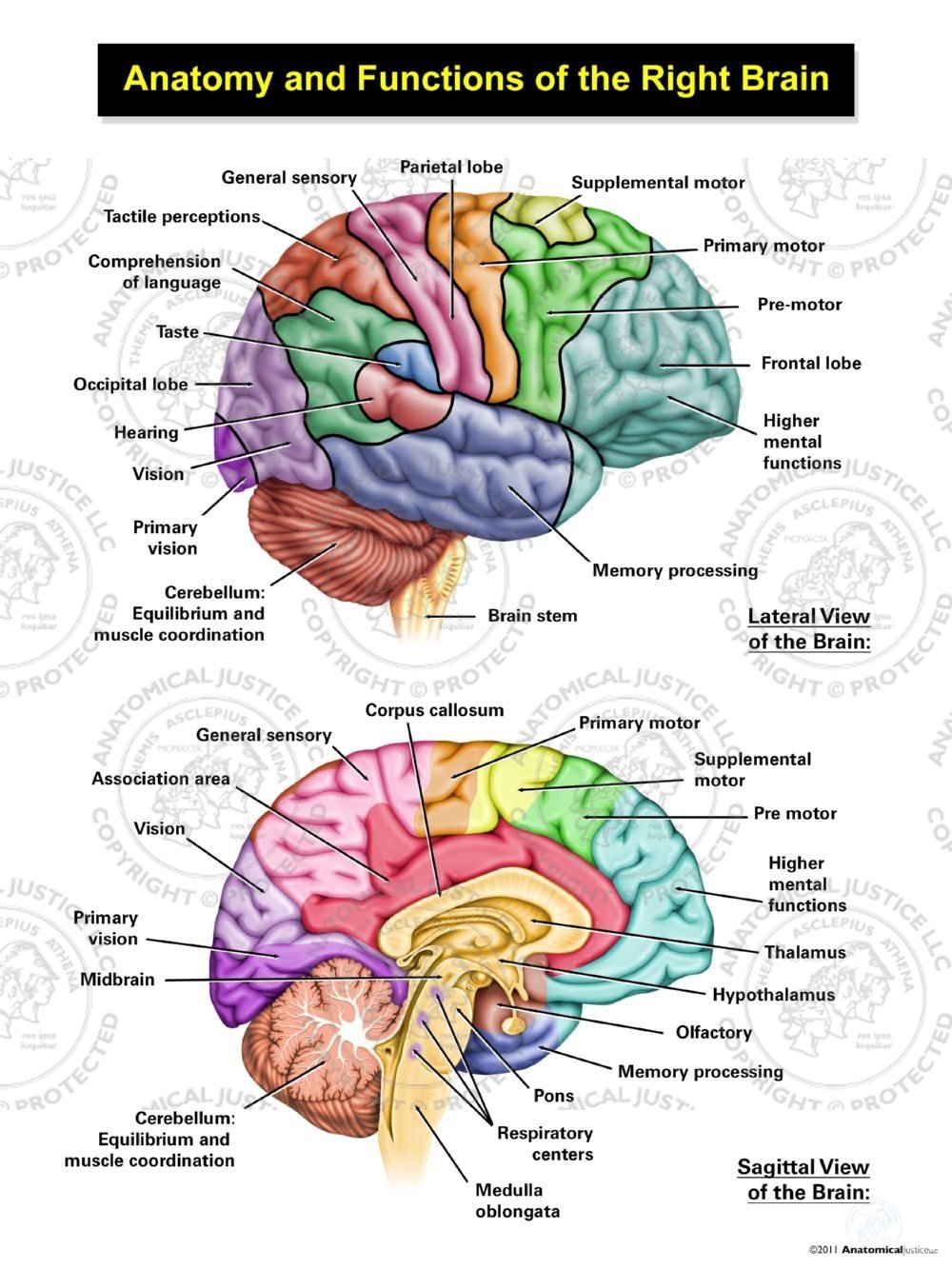
Anatomy and Functions of the Right Brain | Anatomical Justice from anatomicaljustice.com Physiological method of choice to record the electrical activity. The interface combines electroencephalography (eeg) to record brain signals and transcranial magnetic stimulation (tms) to deliver information noninvasively to the brain. Neuroscientists have attempted to break down the different brain parts to explain its structure. Brain and spinal cord, gross. Try some of these examples. Instantly view scientific and medical imaging data in 3d using xtk. The brain is also divided into several lobes: Brain, coronal section, basal ganglia, close view, gross.
Neuroscientists have attempted to break down the different brain parts to explain its structure.
Moreover, brainnet viewer draws the brain surface, nodes and edges in sequence and displays brain networks in multiple views, as required by the user. By their very nature, such experiments tacitly encourage a reflexive view of brain function. Any of a long list of feelings a person can have such as joy, anger and love. External parts of the human brain. The brain is controlling all of these things and a lot more. Probe is pointing to the left olfactory bulb (brain is shown from an anterior view, held upside down); The figure can be manipulated with certain interaction functions to display more detailed information. This module is a comprehensive and affordable learning tool for medical students and residents and especially for neuroradiologists and radiation oncologists. The human brain is the command center for the human nervous system. Study the same and answer the questions that follows: Researchers are breaching the boundaries of the mind, moving information in and out and across space and time. It controls our muscle movements, the secretions of our glands, and even our breathing and internal. It is divided into two cerebral hemispheres.
This module is a comprehensive and affordable learning tool for medical students and residents and especially for neuroradiologists and radiation oncologists. Electrical signals fed back to the brain mimicked the sense of touch. The brain is one of the most complex and magnificent organs in the human body. Neuroscientists have attempted to break down the different brain parts to explain its structure. It is located in the head, usually close to the sensory organs for senses such as vision.
Brain | Anatomy and Physiology I from s3-us-west-2.amazonaws.com This sulcus is an oblique sulcus on the lateral aspect of the brain. Your brain is so powerful and so diverse, it's a big task to name all its different functions. Let's let our imagination loose and chart the scientifically reasonable, machine learning enabled applications that go beyond the matrix trilogy. In an experiment, extract of brain tissue was subjected to differential centrifugation. It is located in the head, usually close to the sensory organs for senses such as vision. It separates the temporal lobe from the frontal lobe. External parts of the human brain. Researchers are breaching the boundaries of the mind, moving information in and out and across space and time.
Monkeys in a recent study used their brains to control a virtual arm and manipulate virtual objects.
A medical illustration of the human brain from 'quain's elements of anatomy, eighth edition, vol.ii' (by william sharpey md, lld, frs l&e, allen thomson, md, lld, frs l&e, and edward albert schafer) depicts the right half of. Each point of view provides an altered perspective of the brain that changes the appearance of the major divisions, landmarks, and structures. Probe is pointing to the left olfactory bulb (brain is shown from an anterior view, held upside down); This sulcus is an oblique sulcus on the lateral aspect of the brain. The brain is also divided into several lobes: It is located in the head, usually close to the sensory organs for senses such as vision. It separates the temporal lobe from the frontal lobe. Brain and spinal cord, gross. One promising strategy for enabling such a system, denoted neuralnanorobotics may also enable a b/ci with controlled connectivity between neural activity and external data storage and processing, via the. Instantly view scientific and medical imaging data in 3d using xtk. It is the most complex organ in a vertebrate's body. A brain is an organ that serves as the center of the nervous system in all vertebrate and most invertebrate animals. If you look at a cutaway view of the brain, you see that the cortical area above the corpus callosum is divided by a.
You have just read the article entitled External View Of Brain / Lab Practical Exam - Biology 255 with Carlson at Luther ... - It provides access to an atlas and to images in axial planes, allowing the user to learn.. You can also bookmark this page with the URL : https://allpendqit.blogspot.com/2021/06/external-view-of-brain-lab-practical.html
Share Awesome
Belum ada Komentar untuk "External View Of Brain / Lab Practical Exam - Biology 255 with Carlson at Luther ... - It provides access to an atlas and to images in axial planes, allowing the user to learn."


Belum ada Komentar untuk "External View Of Brain / Lab Practical Exam - Biology 255 with Carlson at Luther ... - It provides access to an atlas and to images in axial planes, allowing the user to learn."
Posting Komentar